|
OUR MODELS:
The CBV lab currently uses two systems as eukaryotic models: the mouse model and cultured cells (primary, transformed and immortalized). We study influenza A viruses. Our model virus of choice, due to availability of reagents and a powerful reverse genetics system, is influenza A/Puerto Rico/8/34. Note: No model in biology is perfect and must be chosen according to the question to be addressed. Tissue cultured cells have provided a lot of information on viral requirements for host cell components but its use is associated with a lot of problems. Most cells have been immortalized or are transformed and are very different from the primary cells that the virus normally infects. However, if we think that immortalized cells are resistant to a huge array of stimuli, any alteration provoked by a virus to such equilibrium is actually very meaningful. Still, scientific studies could and should be complemented with other models, either in vivo or in vitro. OUR TECHNIQUES:
The CBV lab uses a diversified set of multidisciplinary approaches to molecular biology, biochemistry, and microscopy to unravel influenza A interactions with its host. We highlight a few of the techniques our lab is proudly developing and employing in our ongoing projects: MICROSCOPY BASED APPROACHES:
Microscopy based approaches are powerful tools to observe host-pathogen interactions and alterations to cellular structures during the course of infection. Therefore, we have been developing and ameliorating the following techniques: - CORRELATIVE LIGHT AND ELECTRON MICROSCOPY:
- LIVE CELL IMAGING
- SUPER-RESOLUTION MICROSCOPY -dSTORM
BIOCHEMICAL APPROACHES:
The CBV lab has implemented a system (developed by others) to artificially target proteins to the mitochondria, thus using the cell as a test tube. If a protein is sufficient to recruit a factor in its normal cellular setup, it will be able to do so when the protein location has been changed to the mitochondria, altering their normal intracellular distribution to this organelle. |
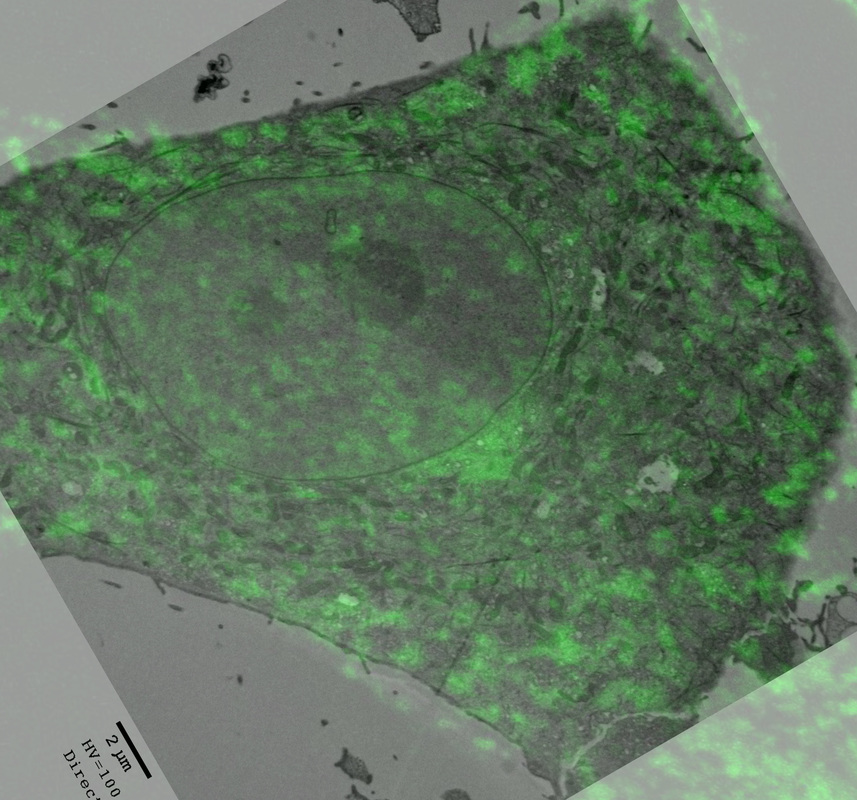
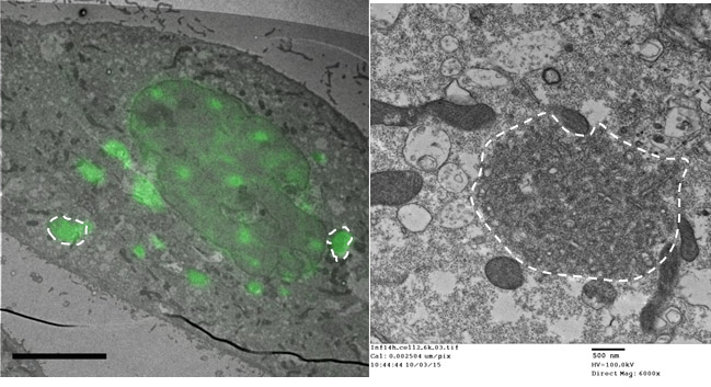
Correlative light an electron microscopy. An example showing correlation between Rab11 protein and aggregation of host vesicles.

Standard electron microscopy image showing a small part of an influenza A virus infected cell.
|
- CELL BIOLOGY OF VIRAL INFECTION lab -
TOOLS
Create a free web site with Weebly