CBV lab
  • Research
  • RESEARCH ERC CoG LOFlu
  • Facts on influenza A virus
  • Our Tools
  • Publications
  • People
  • Photos and news
  • Useful Links
  • Contact
  • Research
  • RESEARCH ERC CoG LOFlu
  • Facts on influenza A virus
  • Our Tools
  • Publications
  • People
  • Photos and news
  • Useful Links
  • Contact
CBV lab

CELL BIOLOGY OF VIRAL INFECTION LAB

REsearch 


​APRIL 2023
​
WE'RE HIRING!

OPENING OF INTERNATIONAL TENDER PROCEDURE FOR HIRING A POSTDOCTORAL RESEARCHER WITHIN THE FRAMEWORK OF THE PROJECT “ERC-2020-COG – LOFLU - 101001521”, DESIGNATED BY “CONTROLLING INFLUENZA A VIRUS LIQUID ORGANELLES”
MISSION STATEMENT:

    The Cell Biology of Viral Infection Research Lab at the Instituto Gulbenkian de Ciência (IGC) studies the interplay between the host and influenza A virus. 
    
    On one hand, we aim to identify the cellular processes that the virus utilizes to promote its own replication and viral assembly. The lab has a grown a particular interest for understanding the formation and material properties of the viral inclusions formed by influenza A virus in the cytosol. Viral inclusions are membraneless organelles that exhibit liquid properties. These structures arise at late stages of infection an accummulate progeny viral RNA. Our current model is that the segmented genome of influenza A virus - that is composed of eight distinct RNA segments - forms a complex in these structures before reaching the plasma membrane where the virion assembles, buds before being released from the cell. By doing so, we help to better define the still incomplete picture of the viral "life cycle", understand how the virus modulates host cell machinery and investigate whether viral inclusions could be specifically targeted to block infection.

    On the other hand, we investigate mechanisms of host and influenza A determinats of virulence. Using this strategy, we study the innate immunity of the host against influenza A virus whilst also determining the mechanisms the virus has developed to escape them.


Note: The image in the header (and the one in the bottom right panel) might seem very strange to you, but it is something we are very proud of. It is an electron microscopy image correlated with fluorescence of an influenza A virus-infected cell expressing the viral protein nucleoprotein that has been tagged with the green fluorescent protein (GFP). Hence the "green" in the figure shows the intracellular location of the nucleoprotein. It is mostly localized in the nucleus (bottom right, big circle) where it is excluded from the nucleoli (the unstained small holes in a bigger green circle). Outside of the green circle is the cytoplasm of the cell where one can detect the opposite coloring system: the majority of the space is unstained but in some places, green circles are detected. These green circles contain de novo-synthesised viral genome on their way to form progeny virions.
Picture
Picture
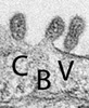
Correlative light and electron microscopy of an influenza A virus infected lung epithelia cell. See explanation on the left (in green).

funding:

Picture
Picture
Picture
Picture
Picture
Picture
                                      ® Maria João Amorim 2022

Create a free web site with Weebly